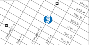
133 E. 58th St., Suite 411
New York, NY
212-688-5770
Sunday
May032009

Hip Pain in Running

Although not injured as often as the knee, leg, or ankle and foot, the hip area is still commonly injured in distance runners. Both veteran an rookie runners alike often experience pain in the hip area.

It is important to understand exactly what, anatomically, the hip area is comprised of, as there are many different structures that can cause pain in the area of the hip. In fact, many runners describe pain as coming from what they think is their hip when the pain is actually coming from other areas nearby, such as the lower back and the rest of the pelvis.

The hip is a joint made by the meeting of the femur, or thigh bone, and part of the ilium, or hip bone. When taken together with the rest of the ilum, the sacrum, and the coccyx and the muscles that attach to these bones, the whole area is known as the pelvic area. The joints between the sacrum and each ilium are known as the sacroiliac joints, one on each side. The major muscles to be concerned with in the hip and pelvic are named below in discussing the specific area of injury.

Functionally, the hip, of course, is subject to impact forces as it plays its role in the running gait. The joint is involved in absorbing the impact of landing, and in the push off and extension of the leg as we propel ourselves forward. These functions cause a lot of stress to be applied to the joint, and if the body cannot handle this stress, overuse injury to the hip will occur. As in all running overuse injuries, the body’s ability to handle the stress of running will be decreased by several factors, such as running too much, too far, and too fast for your training level, not enough recovery time, worn out shoes, and poor nutrition. Also, biomechanical factors intrinsic to your body, such as overpronation, muscle strength imbalances, malignment, leg length inequality, and lack of range of motion/flexibility will decrease the body’s ability to absorb the stress of running without injury. Fortunately, the body does not break down “all at once” (although it may feel that way after a tough race or long run). First to go are usually the “soft-tissues“, meaning the muscles, fascia (which lines the muscles in certain locations), tendons (which attach muscle to bone), bursae (fluid filled sacs that provide gliding of tendon tissue over underlying bone) and ligaments (which attach bone to bone). Also, nerves can be irritated or entrapped. If attention is not paid to these soft-tissues, then the joint and bone tissues will be put at risk for injuries such as degenerative arthritis, hip impingement, labrum tears, and stress fractures, which can be harder to treat. This article will focus on the soft-tissue injuries due to space considerations.

The muscles of the hip must be strong and flexible to give the joint its proper range of motion to do its job. Problems will occur when, because of the factors discussed above, the muscles and/or fascia gradually tighten and lose their normal length. The individual muscle fibers develop adhesions between each other. This change in the muscle itself can be the injury and cause pain upon use. If not released, eventually the tendons, especially where they meet the muscle and where they attach to the bone, can also tighten, swell and weaken. They will not have the strength to do their job. Also, the bursa can get inflamed, but usually only if you totally ignore the pain for weeks or run way longer than you are used to in one run.

What is the first symptom of hip muscle, tendon, or bursa injury? Pain may start as an dull ache during a run or after, although it can be sharper in some muscles or if you have been ignoring the initial pain, and the injury is progressing past the initial stages. Most commonly involved in running are the hip abductors, the gluteus medius and minimus, and the tensor fascia latae (TFL), which is also involved in iliotibial band syndrome (ITBS). (ITBS may be felt at the hip or knee). Also commonly involved are the piriformis and small hip rotator muscles, located just below the gluteals, and the hip extensors (the gluteus maximus and hamstrings). Pain from any of these muscles can be felt at the upper buttock, outer hip, and/or down the outer or back of the thigh. The pain down the thigh is called referred pain, and can be confused with sciatic pain. Next most common are the hip flexors (psoas, sartorius, and rectus femoris), which cause pain in the front of the hip joint and down the front of the thigh and/or groin area. And finally are the adductors, which cause pain in the front and inner thigh,. If your injury is in it’s earliest stages, self-treatment may be helpful. Self-treatment of any of these muscles should start with gentle self-stretching, “rolling” the tight muscle/fascia area on a tennis ball or foam roller, applying ice to the involved areas, and possibly not running for a few days, depending on the severity of symptoms: If you are having pain during of after a run in the muscles that does not go away after two days rest and self-treatment, more rest may be needed. Non-impact cross-training such as the elliptical, cycling, or swimming may be OK. If after a week of rest, pain is still present with daily activities or returns in the same way after a run, professional treatment should be sought.

Professional treatment involves a thorough history and exam to make sure there already is not a joint or bone problem, or a complete muscle tear. The whole lower extremity, from the foot to the lower back, should be examined, and any doctor or therapist you see should be a specialist in running injuries and include a visual or videotape analysis of your walking and running form when the pain subsides enough. This analysis is essential to prevent the injury from reoccurring as it can help identify the predisposing intrinsic factors. If the injury is diagnosed as muscle or muscle and tendon, or also the bursa is involved, manual soft-tissue treatments such as A.R.T. (Active Release Technique), should be used to release the tightened muscle and fascial tissue that is causing the pain. In A.R.T., the doctor or therapist uses his or her hands to contact the tightened area, and either the doctor or patient moves the affected body part to create tension which releases the restriction in the muscle. Treatment will take more visits the longer the injury has been there, because the soft-tissues will be more damaged, or “fibrotic”. Once the muscle and fascia is released, stretches can begin to keep it released. If the area is too tight or swollen for manual treatments, physical modalites such as electric stimulation and/or ultrasound may have to be used first until the area can be treated manually.

The next step in treatment is to correct the training errors and biomechanical deficits discovered throughout the body that caused the injury. The most common general pattern of muscle imbalance related to hip injury is weakness or inhibition of the hip abductors and hip extensors, and tightness of the hip adductors and hip flexors. Other muscle imbalances, such as a weak core, should be addressed as well. Generally, to isolate the weak/inhibited muscles, floor exercises and non-weight bearing exercises involving bands, ankle weights, or machines are first used, progressing to standing exercises with multiple joint motion, such as squats and lunges. Balance exercises on unstable surfaces such as the wobble board, stability ball, and half foam roller are then added, and finally impact activities like plyometric drills (bounding, hopping, and box jumps) are sometimes done. Functional and anatomical leg-length inequality can be corrected by spinal adjustments and heel lifts, respectively. Overpronation secondary to muscle imbalance is corrected with stretching and strengthening, usually of the deep calf muscles, and primary overpronation is corrected with orthotics. Running, starting with even just 10 minutes, then can begin, preferably on soft surfaces such as the reservoir. Resuming running before completing a rehabilitation program invites risk of re-injury, even if you can run pain-free after the soft-tissue treatments! Finally, to keep the injury from reoccurring, as well as to keep your body at it’s highest level of preparedness for running, and maintain longevity in the sport of running, a maintenance, or “prehabilitative” program of self-care is essential! To keep the muscles and other soft-tissues strong and pliable enough to absorb the impact of running, one can continue some of the key rehab exercises, plus use tennis ball/foam-rolling, active stretching pre and post runs, yoga, and strength training including core and upper body postural exercises. Also, it is wise to pay attention to your running form/technique and have a proper training plan for your ability level.

Saturday
Dec082007

Hamstring Injury: Tight 'strings or loose ‘strings? Keeping your Hamstrings Healthy

The hamstrings are a group of muscles which can be injured in all types of sports, and running is no exception. In fact, many runners have hamstring problems and often do not get proper care. Here is a summary of how the hamstrings function, how they get injured, and what to do to prevent and treat hamstring injury.

The hamstrings are actually two groups of muscles which run from the pelvis to the upper leg, on the back, or posterior side of the thigh. The inner, or medial group is made of two muscles, the semimembranosus and semitendinosis, and the outer, or lateral group is made up of one muscle, the biceps femoris. The biceps femoris actually has two parts, or "heads", a long and a short, which can be seen anatomically as two separate muscles. Except for the short head of the lateral hamstring muscle, the hamstrings all originate at the ischial tuberosity of the pelvis, or sit bone. The short head originates at the lateral femur. The semimembranosus inserts, or ends, at the medial, (posterior) back side of the upper tibia, the semitendinosis at the medial, more anterior surface of the tibia, and the two parts of the biceps femoris insert at the lateral side of the fibula. Tendons attach the muscles to the bones, and can be injured as well.

The hamstrings help us move as in running and walking. Looking at them anatomically, they create this movement because they extend the hip and thigh, and they also help bend the lower leg on the upper leg. Throughout the stride they are either contracting or lengthening according to the changing positions of the hip and knee. This function is complex, but we can identify the stride postions that create the greatest risk of injury, as described in the next paragraph. Also, the hamstrings have a function when we are just standing still- they help us remain upright against gravity by exerting a backward pull on the pelvis. This is an 'antigravity' function, and is important for proper posture.

Most muscle injury such as in the hamstrings can be classified according to how it occurs, as either acute (sudden) or chronic (over time). The first type, an acute hamstring injury, can occur during the rapid stretching (lengthening) of the muscle, or during the rapid contraction (shortening) of the muscle. The part of the stride when rapid stretching occurs and injury is possible is when the leg swings foward near the end of the stride. During this time, we are flexing the thigh and simultaneously extending knee, stretching the hamstring from both ends, and tearing can occur. The part of the stride when rapid contraction occurs and injury is possible is during late push off. During this time, the hamstrings rapidly change from stabilizing the knee during the support (foot on the ground) phase to extending the hip as the knee flexes. The lateral hamstring is most likely injured at this time. In either case, as the injury occurs you feel immediate pain, usually described as a "pulled muscle". The location of the tearing is usually high in the thigh, near the where the muscle meets the tendon, or slightly below, towards the middle of the muscle. The injured area will feel sore to the touch, and the torn fibers may present themselves as a lump of tissue. The area may or may not be swollen. The tears can be graded as mild (grade 1), moderate (grade2), or severe (grade 3) depending on the amount of tearing. Fortunately for distance runners, these sudden types of injury usually occur during sprinting sports which require sudden, explosive movements, such as football, baseball and the track and field sprints. One exception, however, is when a runner finishes a distance race with a sprint, which can suddenly overload the hamstrings, which are usually tight and fatigued by that time in a race. In that case the injury will resemble more of an acute tear.

The second type of injury, the chronic one, is the more common one for runners, especially in high mileage or older runners who have been running for years. The injury usually occurs more gradually. With distance running, slight muscle tightness in the back of the knee, where the hamstrings insert, may not cause pain. But when we use a muscle often over and over for an extended period, as in distance running, the individual fibers can start to get "stuck" to each other. Upon manual examination, painful "knots" or adhesions can be felt by hand. If not treated, they then can lead to shortening of the muscle lengthwise- muscle tightness. With this tightness in place, the muscle will not lengthen properly or contract as strongly when exercised. This can eventually cause pain when enough fibers become involved. In this situation the location of the tightness is usually very high or low in the muscle, or the tendon itself can have the scar tissue. This muscle fiber tightening usually occurs either over the course of one workout or over days or weeks. Examples of immediate causes are a longer run than you are used to, or a faster speed session. It is important to note that an area of chronic tightness and/or weakness is often more susceptible to also suffering an acute tear during an end of a race sprint as described above.

Before a diagnosis of chronic hamstring injury is reached, other conditions such as sciatic nerve irritation or a pelvis stress fracture should always be ruled out. However, two injuries can exist at once, and can be related, so the hamstrings should be checked anyway if the other pain sources are found.

There are several reasons the hamstings are injured in sports. First, there is muscle imbalance, meaning one muscle is stronger than its opposite functioning muscle. In this case, the quadriceps, which extend the knee and flex the hip, are the opposing muscle group to the hamstrings, which flex the knee and extend the hip as mentioned above. If the quadriceps are overly developed in comparison to the hamstrings, the hamstrings must work harder to pull against the quadriceps, and can strain or tear if they are not strong enough. This is often the case in sprinting injury. The other common imbalance is the hip flexors being tighter than the upper hamstrings and gluteus maximus, which are the hip extensors. This is more often the case in distance running. In this case the upper hamstring will have to contract harder and can gradually tighten or become strained.

The next predisposing factor is adhesion formation, in the hamstrings and also in related muscles. As noted above, the adhesions will cause tightness and pain, with the tightness leading to more pain and so on. If untreated, then with the decreased elasticity there is even less range of motion, and the uninjured fibers will have to work harder, causing them to tighten and form new adhesions. This makes the chronic injury more likely to keep occuring. Also, muscular adhesions in the related hip muscles (gluteals, piriformis) can refer pain to the hamstrings. This can cause a feeling of hamstring strain, when none is really present, or both the hamstrings and the hip muscles can have adhesions. Also, the sciatic nerve can be "caught up" in hamstring adhesions, or further up above in the hip muscles, and either problem can further tighten the hamstring or simulate the feeling of a strain.

The third factor is pelvic alignment problems. Many people have postural distortion in the form of a lumbar spine or pelvic rotation and tilting, which causes an apparent "short leg" as one side is higher or lower than the other. Everyday activities may or may not be painful, but when subjected to the repetitive stresses of training for a marathon, the result can be muscle injury. The hamstrings can be overstretched on one side in this case, often the "long"side, and the gradual or acute type of hamstring injury then can occur.

Treatment of hamstring injury will vary depending on the both the degree of injury and which of the above predisposing factors need to be addressed.

In acute injury, self-treatment will involve ice application, with a compression wrap and elevation, at least once a day. Ceasing running for at least 2 weeks even in a mild, grade 1 tear must be done as, training through the injury will not help, and recovery must come first. Cross-training with activities that keep the range of motion to the pain-free level can be done, such as the elliptical or stair machines. In grade two tears return to running may take up to 4 or 5 weeks, and in grade three 2 or 3 months. It is important to note that the amount of healing time can vary in different people. Soft-tissue manual therapy such as active release technique must be done to minimize scar tissue formation and adhesions, and can greatly speed up healing in all three grades of tears. If the adhesions are not broken up, then the tear will heal with less flexible tissue than normal, healthy tissue and re-injury or the chronic tightness will occur. In all cases, return to running should be gradual and include an extended warm-up period, and speed training should not be done until easy and mid-paced runs are completely pain-free. Third degree tears may require surgical intervention if the tendon is fully pulled off the hip bone. This is rare, but if it occurs immediate surgery is neccesary for proper healing.

In the chronic injury, the tightness must be treated by removing/reducing the adhesions in the muscle or where the muscle meets the tendon. Again, manual therapy will release the adhesions. The related muscles, such as the gluteals, piriformis, and hip flexors and also the adductors and calf muscles also should be checked. This will remove the adhesions as a pre-disposing factor. Also, the "referred hamstring pain" and sciatic nerve adhesion syndromes described above can be fixed by the active release technique.

In both the acute or chronic injury types, strengthening exercises should be done, which can help correct any predisposing muscle imbalance, but not until the manual therapy has released most of the muscle adhesions. First are gentle range of motion exercises, progressing to isometric contractions (no joint movement) and then full-range of motion with light resistance at first. Eventually, the hamstrings should be strengthened during the lengthening movement (which is when injury often occurs), which means squats, lunges, and even backwards running for short distances (40-50 yard repeats in a safe area). Plyometric, or jumping exercises can help ready the hamstrings for explosive movements such as in the final kick of a race, plus stengthen the muscle-tendon junction, where the chronic tightness/weakness often occurs. Exercises on the stability ball are also excellent for strengthening the core as the hamstrings are worked.

The last predisposing factor, lumbar and pelvic alignment problems, are fixed by spinal manipulation of the rotated lumbar vertebrae or ilium (hip bones). This will correct a "functional short leg". Anatomical differences in leg length can be treated with heel lifts, and overpronation, which can lower one side of the pelvis, can be fixed with orthotics. Removing lumbar joint restrictions also will help with uneven torso rotation which can cause an uneven stride, which can contribute to tightening of the hamstrings.

Hamstring injuries can be very stubborn and are often difficult to treat, but if the above factors are addressed, the chances of re-injuring an acute injury or struggling with the chronic one will be diminished.

Thursday
May102007

Achilles Tendon Injury: Avoiding an 'Achilles Heel'

According to ancient Greek mythology, Achilles, the warrior, was dipped by his mother in the magic waters of the river Styx to make him immortal. However, she held him by the heel, which remained dry. The legend says this left the area vulnerable. Subsequently, he was killed in the Trojan War when Paris shot an arrow into Achilles’s heel. But his name lived on, as the tendon that attaches your calf muscle to the heel bone (calcaneus) was later named for him. In running and many other sports, this area can be just as susceptible as it was for Achilles. This article will explain why the achilles tendon is one of the most commonly injured tendons, how to recognize early signs of injury, and what the proper treatment is.

The muscles to which the achilles tendon attaches are the gastrocnemius and soleus. When these muscles contract during the running stride, they function as plantar flexors (downward motion of the foot), which gives us propulsion forward. When they stretch during the running stride, these muscles help in absorbing the impact of landing as the foot comes towards the knee. Also, since the gastrocnemius crosses the knee joint, when it contracts it helps in flexing your knee during the stride. The forces these muscles generate will affect and be affected by the health of your achilles tendon.

With the above anatomy and biomechanical information in mind, it is easy to see why the achilles is often injured. Common injuries are partial or complete rupture and tendinosis (scar tissue development). Until recently, overuse injuries to tendons such as the achilles were thought to involve inflammation. However, research has shown that there are no inflammatory cells present in most injured tendons. Instead, there are fibroblast cells, which form “scar tissue.” This scar tissue forms when the stress placed on the tendon is too great, causing degenerative microtears. Although the tears are “healed” with this new scar tissue, it is, unfortunately, not as flexible or resilient as the original tissue. Additionally, the most common area of injury to the tendon is about 2 cm above its attachment at the heel. It is thought that this area has less circulation and therefore is prone to the mircrotears.

Injury to the Achilles tendon can either happen suddenly, as in a rupture,or can occur gradually, as with tendinosis, which is the usual case in running. In the sudden injury scenario, recreational athletes often injure an already tightened or weakened calf when it is suddenly stretched or contracted past its normal limits, causing the tear. Although tears can occur in endurance athletes, the common scenario for a tear is a middle-aged person who has gradually lost flexibility and developed adhesions in the calf muscles and around the tendon, probably from years of sitting without regular stretching, and who goes out and plays tennis or softball for the first time in months or years. He or she then stresses muscles and tendon past their current limits, and a tear occurs. So even though the injury may seem sudden, the predisposing factor of decreased calf flexibility was probably present for some time. Likewise, in running and other endurance sports, a loss of calf flexibility and calf muscle/tendon adhesions are also to blame for cases of tendinosis.

To prevent overuse injury to the achilles, addressing the factors leading to tightness and possible scar tissue formation is necessary. Muscle tightness can be a major factor, and two areas are often the culprit: one is the calf, or back of the leg, as mentioned above, and the other is the adductor, or inner thigh muscles. Regular stretching and strengthening of these muscles (exercises described below) will help preserve normal foot motion and prevent overpronation. This is important to prevent, because overpronation, which is the ankle bone “rolling inward” at a greater than normal amount or speed, can overstretch the achilles. Overstretching causes the tendonosis by stressing the tendon and causing the tendon microtears and adhesions (the scar tissue). Tight calf muscles can cause a compensatory overpronation, to make up for the lack of leg bend at the ankle by flattening the arch more than normal. Likewise, tightness in the adductors can lead to the thigh moving inward while running, which then can cause our foot to turn outward, pronating excessively to compensate and keep us moving forward.Overpronation can also exist without any muscle tightness or imbalances, and lead to injury in the same way. Orthotics will correct the overpronation. Calf weakness can also be a factor, as lack of strength will cause overstretching of the achilles. Finally, training errors, such as doing too much speedwork or rapidly increasing training volume, can be causative. So can wearing worn out shoes, which can cause overstretching and tightness which will lead to injury. Unfortunately, often a runner often will only learn about these preventative measures once injured. The good news is that with proper treatment, even the worst cases can be resolved; however the earlier treatment starts, the better.

The type of treatment needed will depend on the stage of the injury. Depending on the severity of injury when treatment is initiated, at least one and as many as three weeks off from running are usually necessary. In the earlier stages of achilles injury, there is usually calf muscle and achilles tightness which causes tightness and pain with either contraction (push off) or stretching. This tightness must be addressed by techniques such as Active Release Technique, which reduces the muscle adhesions and increases flexibility. This reduces stress on the tendon, and often soreness there will decrease without direct treatment of the tendon. If the injury has progressed to swelling around the tendon, ice massage and electrical stimulation will help decrease the edema. This swelling is often felt as a “creaking” feeling when holding the tendon lightly and bending the foot up and down. Or, if the injury involves scar tissue formation, as in the athlete with a history of repeated episodes, cross-friction massage will be needed. Cross-friction works to increase the elasticity of the tendon by “breaking up” the scar tissue and restoring flexibility. Also, if there is restriction in motion of the ankle joint, manipulation of the ankle can help relieve stress on the Achilles. In this acute stage, taping for support and using heel lifts (in both shoes) to decrease stretch on the tendon will speed healing. All of these measures should greatly reduce the severity of the injury within three to six treatments, in most cases.

Once the acute pain stage is over—which means there is only mild to moderate tenderness over the tendon and no more pain on push off—the other factors named above must be addressed. This includes Active Release of any muscle tightness further up from the injury, such as in the adductors. The whole “kinetic chain”, from the leg to the hips, lower back, and even upper back, should be evaluated by analysis of the running form and treatment should address the imbalances found. Orthotics should be prescribed if overpronation is found, and worn-out shoes should be replaced. Stretching should be done the “active” way, by using a towel or stretching cord and contracting the muscles which oppose the calf. While seated with the legs out in front of you, actively contract the muscles in the front of your leg while using the towel or rope to assist near the end of the stretch. Hold for two to three seconds and repeat five to ten times. No pain should be felt at the tendon with these stretches. Stretches for the adductors or any other tight muscles, of course, should also be done. Strengthening exercises should include calf raises, with emphasis on the lowering, or “negative” part of the exercise. Standing on a step, raise up on the toes with both legs, and then lower slowly using a count of five to ten seconds. Repeat for ten repetitions and work up to three sets, after which weights can be added by using a calf-raise machine. Although it is doubtful that these guidelines could have saved the warrior Achilles, they could help save your running.

Wednesday
Nov082006

Ankle Pain and Running, Part 2

In part 1 of this article, I discussed how hidden ankle problems, caused by “scar tissue,” can affect the kinetic chain and cause injury elsewhere, such as a stubborn hamstring strain, knee pain, or even back pain. I am not referring to a brand-new, acute injury, but dysfunction that may be present from an injury a long time ago. Here in Part 2, how to find out if you have hidden ankle injury will be discussed, as well treatment and some of the exercises to do to fix it and get you healthy again.

The first step is to test your ankle for the scar tissue. One way to do this is to self-test your ankle for tenderness, restricted range of motion/muscle tightness, and decreased balance ability. As presented in part 1, there are two main groups of ligaments in the ankle, the lateral (outer) group, and the medial (inner) group. It may help to look at the diagrams from part 1. To test the lateral ligaments, start by locating the end of your fibula, which is marked by a thickening of bone called the lateral malleolus. Then run your fingers forward about half an inch, and you will be on the most commonly injured ankle ligament, the anterior talofibular ligament. Often only mild pressure is necessary to note some tenderness. Compare both ankles simultaneously. If one side is tenderer, there may be a problem with the ligament. For the remaining two lateral ligaments, you have to move your fingers straight downward and slightly backward from the lateral malleolus and test for tenderness there (calcanealfibular ligament), and also move straight backward from the malleoulus about half an inch and test there (posterior talofibular ligament). For the medial ligaments, find the medial malleolus, which is the thickened bone on the end of the inner tibia. If you move your fingers forward half an inch, you will be on the anterior tibiotalar ligament. Slightly below that is the tibionavicular ligament. After checking for tenderness in these, again start at the medial malleolus and move straight down, and you will be on the tibiocalcaneal ligament. Finally, from the malleolus move straight back and slightly downward and you will be on the posterior tibotalar ligament. Again, for all these ligaments, compare left and right, noting any tenderness. Healthy ligaments are not painful to touch, even with relatively deep pressure! It is possible to have problems on both left and right sides. If you have difficulty finding the ligaments it may be necessary to seek help from a knowledgeable sports medicine professional.

The next step is to test for reduced range of motion. Two tests can be done. Don’t worry; no major anatomical knowledge is necessary for this one. Sit on the floor, with your legs straight out in front of you, and no shoes on. The first test is to just stretch your calf muscles by actively bringing your foot straight up towards your shin. Do this on the left and right simultaneously, looking for a decrease in the range of motion. You should be able to bring the foot up 20-30 degrees from the rest position. If you use a rope to pull even further, up to 45 degrees may be possible. If there is restriction, it may be in the calf muscles behind you leg, or in the ankle joint itself, or both. Tightness in the ankle joint may mean the ligaments have scar tissue that needs to be released, as the tissue is restricting the ankle bone (talus) from moving. If you only feel muscle tightness, then you may only have shortened calf muscles on that side. If the muscle has been tight for a long time or has had previous injury, it may also have scar tissue that needs to be released. The second test involves putting your foot on the ground, knees bent, and sliding them away from you. Do not let your heels come off the floor. One side may feel more limited. If it does, again it can be either in the joint or the muscles. In this test, the muscles in front of the leg are being tested.

The third self-test is for decreased balance ability. Standing barefoot in a doorway, bend one leg at the knee. You are testing the leg on the ground. If your balance is proper, you should be able to stand straight for 30 seconds without picking up on either the inside or outside of your foot and without your hands reaching side to side significantly for the doorway. As mentioned in part 1, scar tissue in the ligaments can alter your balance ability.

Now, if you have found problems, fixing them usually involves a combination of professional treatment and self-treatment with exercises. When I look for this hidden ankle dysfunction in a patient, the same tests mentioned above are part of my exam. I also watch the patient walk and run, to evaluate how the ankle joint is moving in real life. This helps give a proper diagnosis of why the ankle is not working correctly and/or if the ankle is playing a role in injury elsewhere. The scar tissue will be more developed the more severe the original injury was, and the longer the period of immobilization that may have occurred. Even though it is optimum to get any injury treated as early as possible, the scar tissue can still be made more flexible and actually heal to be much more like the original ligament tissue if it is discovered later on.

To do this, treatment involves restoring length to the scar tissue, which then helps restore the range of motion to the ankle joint. This is done by soft-tissue techniques, such as Active Release Technique (ART) and cross-friction massage. In ART, the provider makes a specific finger contact at the site of the scar tissue, and then the ankle is moved to create tension on the ligament as the provider maintains the contact. The contact and tension is in line with the fibers of the ligament. This combination of tension and movement “breaks up” the scar tissue, restoring proper length and strength to the ligament. In cross-friction massage, the scar tissue is broken up by a finger or massage tool contact moving across the fibers of the ligament. All the ligaments, plus the joint capsule, must be evaluated.

Once the scar tissue in is treated, the muscles are checked. Scar tissue in the muscles is often called “adhesions”. ART is very effective at breaking up the muscular adhesions as well. Besides the calf muscles, the rest of the body must be checked. With chronic ankle injury, there often can be a reflex weakening of the gluteus medius, or hip abductor, so it may have adhesions and weakness. Also, the original ankle injury may be related to a nerve problem (sciatic nerve, for example) anywhere from the ankle to the lower back, as nerve entrapment can weaken the leg muscles and pre-dispose to a sprained ankle. Thus the back should also be evaluated and treated as necessary.

Once you have been treated, stretching exercises must be done to maintain the increased range of motion. To stretch the gastrocnemius muscle in the back of the leg, use a towel or rope, just as in the range of motion test described above. Actively move the foot towards the shin, using the rope to assist the last quarter of the stretch, and hold for 2 seconds. To stretch the soleus muscle, which doesn’t cross the knee joint, bend your knee and then use your hands to pull the foot upward. There will not be as much movement in this stretch. To stretch the tibialis anterior in front of the leg, from a sitting position, bend the leg to be stretched and place the leg over the straight leg above the knee. Then take your opposite hand and pull the foot downward gently and holding for 2 seconds. Repeat all of these stretches 5-10 times, attempting to move further each time but not forcing the stretch.

Strengthening exercises include using elastic tubing in two ways. One is to do small movements for 10-60 seconds. Small movements help improve coordination, as well as strength and endurance. Start in the middle of the range of motion of the exercise and move 5-10 degrees in both directions. Speed can be increased as endurance is gained. In the beginning you may only be able to do 10 seconds before fatigue sets in. Do a total of 60 seconds, by building up to 2 x 30 seconds. The other tubing exercise is to do full range of motion for either time or repetitions. Internal rotation, external rotation, plantar flexion and dorsiflexion all can be done for these exercises.

Balance exercises must also be done, to restore the lost proprioception that occurs with chronic ankle injury. First, simply repeat the self-test described above, for a total of 60 seconds standing (2x 30 seconds). Once this is easy, try with the eyes closed. Then, balance boards and rocker boards also can be used.

If hip or lower back/core muscle weakness was found with examination, then strengthening for those areas must be done as well. Once all weak areas have been strengthened and balance ability improved, running drills and plyometric (jumping) exercises for coordination, power and agility can be added. This complete approach, starting with removing soft-tissue restriction, then improving range of motion, strength, and balance, can help the body heal hidden ankle injury and the problems it may lead to along the kinetic chain.

Tuesday
Aug222006

Keeping Your Back On Track, Part 2

Once the relationship between anatomy and lifestyle and their effects on the back has been understood, then steps can be taken to address the physical, chemical and emotional stressors which lead to back injury. Solutions for combating the physical stressors of running will be discussed here. Before I get started, it is important to note that there are non-musculoskeletal causes of back pain, such as referred pain from abdominal or pelvic organ disease. Whenever a patient presents to my office, a thorough history and examination is done to rule these out before treatment begins. If necessary, diagnostic imaging, such as x-rays or MRI, is done. Besides helping in ruling out the non-musculoskeletal causes of back pain, these studies can reveal any bone abnormalities and arthritic conditions, which may play a role in causing back pain. However, it is important to remember that many people with imaging findings of disc degeneration or disc herniation, for example, may not be in constant pain if the functional ability of their muscles is at a high level. The “whole patient is treated, not the x-ray”, which is one reason why so few people need surgery as the solution to their back problem.

In general, addressing the physical stress of running on the lower back involves maintaining the core strength and overall flexibility reviewed in the above information on anatomy. However, there can be differences in the type of lower back pain experienced by beginner runners and veteran runners. Beginners and those who have not been running in a while often have pain from tightness and weakness in the lower back extensor muscles, especially when running hills. Pain may be present only during the run, and may or may not disappear as you warm-up. The solution can be as simple as adapting to running again; as conditioning improves the pain should decrease. However, if you have been really inactive or if pain persists after a few runs, core strengthening and general flexibility work are probably needed.

In veteran runners- especially in marathoners, muscle imbalances can lead to lower back pain and/or sciatic type pains in the buttocks, hips, hamstrings and calves. Distance running tends to tighten these muscles, as do many other activities, such as sitting at work, and after many years of both activities pain can develop. Pain down the legs can be "psuedosciatica", referred from restricted spinal joints of the lower back and/or from "trigger points", which are knots in the muscles of the lower back, hips and legs. However, disc problems can irritate the spinal nerves and cause "true sciatica" with pain anywhere along the path of the sciatic nerve.

In beginners and veterans alike, the type of treatment necessary depends on if the pain and injury is in the acute or chronic stage. For example, a runner might ignore the warning signs of persistent muscle tightness after longer runs, and eventually experience pain, either during or after a run, that is very severe and incapacitating. Treatment will start with reducing or stopping training and applying ice to the area to reduce pain, swelling and spasm. Office treatment will include electric muscle stimulation, ultrasound heat treatments, and chiropractic adjustments, to get the tightened muscles and joints moving again. Next are pain-relief stretches. The exact exercises to be done in cases of acute lower back pain are very specific to each individual’s injury. Some people need "flexion exercises" and some people need "extension exercises" to help mobilize the spine and relieve pain, and this is decided with the help of the doctor. Once the pain relief stage is over, corrective exercises and joint and muscle balancing techniques must be done to prevent re-injury, just as in chronic injury, which is described below. It is important to note that anti-inflammatory medication or steroid injections may help in relieving the acute pain in some cases, but should never be the end-point of treatment, as they do not address the underlying, functional causes and leave one open to re-injury.

Treatment of chronic LBP in runners must address the functional imbalances that cause pain themselves or allow degenerative joint and disc problems to become symptomatic. This involves identifying which muscles are tight and/or weak, and which joints are not moving properly. Common muscle patterns involve tight hip flexors, especially the upper quadriceps and psoas muscle, with weakness in the opposing hip extensors (gluteus maximus and upper hamstring) muscles, or tight lower back extensors with weakness in the opposing lower and deep abdominals. These are some of the vital “core” muscles mentioned in part one. Further down, the lower hamstrings and calf muscles are often tight. Also, the hip abductors (outer hip) are often weak and the adductors (upper, inner thigh) tight. Before the weak muscles can be strengthened, the tight ones must be stretched. Office treatment involves manual release, such as Active Release Techniques, of muscle trigger points and adhesions, which is often essential in restoring proper flexibility and strength. Then, stretching can be done with assistance, as part of office treatment, and by using self-stretching techniques at home, such as active-isolated stretching. As for joint restriction, the lower thoracic and all of the lumbar vertebra, plus the sacroiliac (between the hip bones and sacrum) joints, are often involved. Office treatment includes spinal and extremity adjustments to restore normal joint motion and coordination.

Once normal muscle length is restored, strengthening and stabilization exercises are added, starting with isometric deep abdominal and deep back extensor contractions, and progressing to holding this core tight while performing arm and leg movements, and then to exercises such as standing lunges and squats. Balance exercises and full-body movements such as pull-ups and push-ups are also beneficial, as they strengthen the upper back, which is essential in maintaining an upright posture during long runs.

Other factors that need to be addressed for successful management, if they exist, include correcting overpronation and leg-length inequalities with orthotics and/or heel lifts. And, of course, training errors must be examined, such as too many hills when just starting to run or after a layoff, worn out shoes, only running on pavement, too many long runs in a marathon training plan or not enough recovery between long runs.

This program of treatment will prevent the cycle of minor joint strain and muscle tightening, pain, emotional disturbance, and further physical damage from occurring. It will keep the healthy muscle tissue from being damaged and replaced with fibrotic scar tissue, which is less elastic, weaker and more prone to injury.

Because this information is often learned only after serious injury and training interruption, an earlier understanding and proper intervention may make it easier to keep your back, and your training, on track.

Make an appointment

Email us at ptcompete@gmail.com

Office News & Upcoming Events

Prepare to Compete Injury Prevention Clinics:

2025 Clinics to be announced soon!

Directions

133 E. 58th, Suite 411
New York, NY 10022
Phone: 212-688-5770

© 2012-24 Bochner Chiropractic and Sports Injury Care. All Rights Reserved.